
Increase in intracellular calcium concentration [Ca2+]i triggers a series of cellular events including neurotransmitter release, muscle contraction, and oocyte fertilization (Clapham, 1995; Berridge MJ, 1993). The elevated state of the intracellular calcium level and the response to this elevation varies depending on the agonist and the cell line. It has been reported that the intracellular entry of the calcium-sensitive dye Fura-2 (Grynkiewicz, 1985) leads to changes in the level of [Ca2+]i in subcellular levels in all cell suspensions, adherent cell populations (Lambert D., 1992).
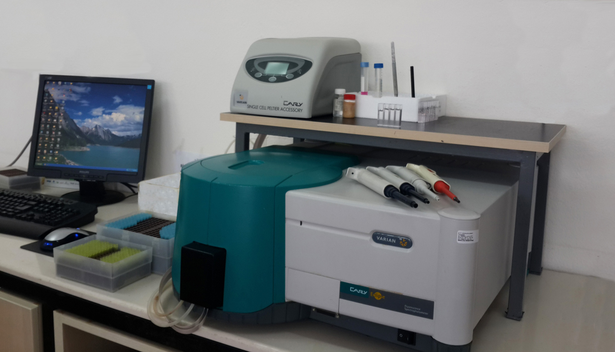
In the nanoscale dimensions, a large number of [Ca2+]i signals within microseconds are used by different cell types in different scales ranging from the [Ca2+]i signal waves in subcellular areas to all cell [Ca2+]i signal waves (Berridge, 2006; Bootman et al., 2001 ). The characteristics of these cellular Ca + 2 signals are due to the expression of tissue-specific Ca2 + transport systems (Berridge et al., 2000). The diversity of cellular [Ca2+]i signaling means that there is not a single technique that can be used to monitor [Ca2+]i changes in all cases.However, fluorescent ratiometric [Ca2+]i signal measurement techniques are widely used as a versatile way to analyze cellular [Ca2+]i signaling responses. Since its first disclosure by Tsien et al. (Tsien et al., 1982), fluorescent [Ca2+]i indicators have been used in the investigation of [Ca2+]i signals in various cell models in different experimental environments. Using appropriate fluorometric meters and appropriate indicators, it is possible to monitor [Ca2+]i signals on live cells without time-lapse, high-rate, including subcellular levels. This section describes practical approaches to the measurement of [Ca2+]i signals by the use of synthetic fluorescent indicators such as Fura-2AM to monitor intracellular [Ca2+]i concentration.
Calcium Signal Course Program
Lecture 1 Preparation of solution (1xPBS, calcium buffer, Calcium Free Buffer etc.)
Lecture 2 Calcium Signaling Analysis-1
Lecture 3 Calcium Signaling Analysis-1
Lecture 4 Caspase 3, Caspase 9 and Apoptosis Analysis
Lecture 5 Analysis of intracellular ROS and mitochondrial depolarization
Lecture 6 Participants Making the Calcium Signaling Analysis-1
Lecture 7 Participants Making the Calcium Signaling Analysis-2
Lecture 8 Participants apoptosis, caspase-3, -9, Making the intracellular ROS and mitochondrial depolarization analysis.
Lecture 9 Calculation of results, statistical analysis, and ready to be presented in the article-Making Drawing Figures
Lecture10 Group discussions and advice